Celleste Image Analysis Software
Coupled with the powerful image acquisition capabilities of the EVOS microscopes, Celleste software allows you to seamlessly capture, process, segment, classify, measure, analyze and share images and data.
Download free trial of Celleste Image Analysis Software
Description |
Single-user License |
Network License* |
Celleste 6 Image Analysis Software |
||
Celleste 6 Image Analysis Software Upgrade** |
||
Celleste Image Analysis Software Network Seat, One additional seat |
NA |
|
*Network licenses includes one seat that can be assigned by the administrator to multiple users across an internal network. Only one user can access a seat at one time. |
||
Base functionality
Celleste 6 software offers powerful tools to segment, count, size, classify and analyze complex images, including machine learning algorithms for advanced image analysis and quantitation. The Celleste 6 Multi Channel Analysis (MCA) tool is based on pre-configured algorithms and analysis templates that have been trained on representative data to optimally segment and classify images from a range of common cell-based assays. Simply choose the app that corresponds to your assay of interest and follow the wizard-based workflow step by step from image to data generation.
Alternatively, use the full suite of powerful tools in Celleste 6 software to customize image analysis to your specific needs, including image adjustments, background correction, alignment and tiling, counting and sizing using powerful segmentation and classification tools, and measurements in up to four dimensions applied automatically over multiple channels, fields, and wells.
Optional modules
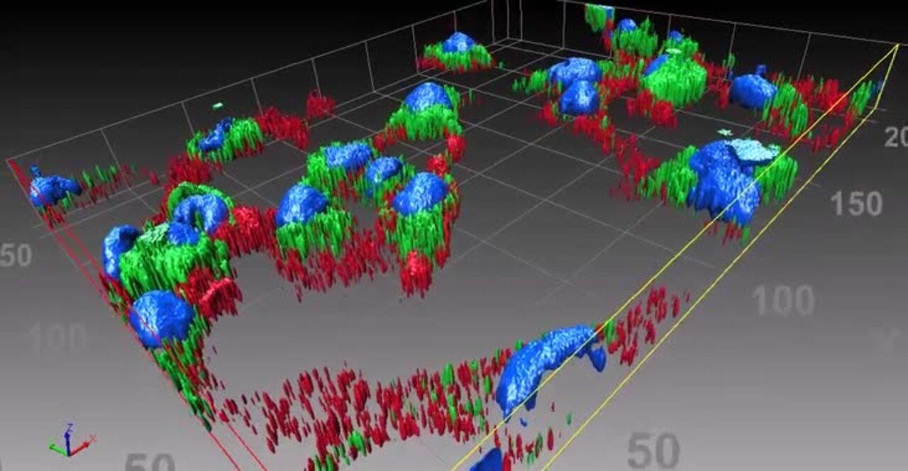
Optional modules for deconvolution, 3D visualization, and 3D analysis allow you to customize the capabilities according to your needs.
Module Description |
Single-user License |
Network License* |
Module Detail |
Celleste 2D Deconvolution |
Improves single-plane image quality (S/N) of cells or tissue slices by clearing background haze (out-of-focus light). |
||
Celleste GPU Acceleration for 2D Deconvolution |
Deconvolves 2D image sets in a fraction of the time by utilizing the graphics processing unit (GPU) of your EVOS PC or workstation |
||
Celleste 3D Deconvolution |
Improves resolution and clarity of thick samples like spheroids, tissue slices, or cells in 3D matrices by deconvolving image Z-stacks. Watch video |
||
Celleste GPU Acceleration for 3D Deconvolution |
Deconvolves 3D image sets in a fraction of the time by utilizing the graphics processing unit (GPU) of an EVOS PC or workstation |
||
Celleste 3D Visualization |
Tools to adjust background and transparency, apply different projection types, and create and enhance 3D movies.
|
||
Celleste 3D Analysis |
Provides guided segmentation tools for complex 3D objects to facilitate identification and measurement |
||
*Network licenses includes one seat that can be assigned by the administrator to multiple users across an internal network. Only one user can access a seat at one time. |
|||
| Before | After |
| Before | After |
Applications
The Celleste 6 image and analysis tools are useful in a broad range of applications, such as cell viability, apoptosis, cell cycle, live/dead cell analysis, transfection efficiency, immunohistochemistry analysis, migration tracking, wound healing, adipogenesis, and cytoskeletal disruption. The Celleste 6 Multi Channel Analysis (MCA) tool is based on pre-configured algorithms and analysis templates that have been trained on representative data to optimally segment and classify images from a range of common cell-based assays. For application notes detailing the use of Celleste software in several of these applications, and references to published papers that have used it, see Resources.
How-to videos
These short videos explain how to accomplish tasks in Celleste software. The Celleste 6 Multi Channel Analysis Tool (MCA) simplifies many of these tasks including among others wound healing, batch processing, colocalization and parent-child), but all of these tools can also be used to analyze images manually.
Celleste base functionality
Celleste optional modules
For Research Use Only. Not for use in diagnostic procedures.
