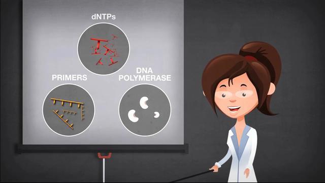
The success of PCR depends on a number of factors, with its reaction components playing critical roles in amplification. Key considerations in setting up the reactions include the following PCR components detailed on this page:

A PCR template for replication can be of any DNA source, such as genomic DNA (gDNA), complementary DNA (cDNA), and plasmid DNA. Nevertheless, the composition or complexity of the DNA contributes to optimal input amounts for PCR amplification. For example, 0.1–1 ng of plasmid DNA is sufficient, while 5–50 ng of gDNA may be required as a starting amount in a 50 µL PCR. Optimal template amounts can also vary based on the type of DNA polymerase used; a DNA polymerase engineered to have higher sensitivity due to affinity to the template would require less input DNA. Optimization of DNA input is important because higher amounts increase the risk of nonspecific amplification whereas lower amounts reduce yields (Figure 1).

Figure 1. Comparison of PCR results with plasmid vs. human gDNA template. The same DNA polymerase was used to amplify a 2 kb target sequence from varying amounts of input DNA under the recommended conditions.
At times, PCR protocols may call for input of DNA in terms of copy number, especially for gDNA. The copy number calculation depends on the number of molecules present, in moles of DNA input. Using Avogadro’s constant (L) and molar mass, copy number can be calculated as:
Copy number = L x number of moles = L x (total mass/molar mass)
The molar mass of a particular DNA strand is determined by its size or total number of bases (i.e., a combination of its length and single-stranded or double-stranded nature). For convenience and simplicity, an online tool is available to calculate copy number from the mass of the input DNA.
In theory, a single copy of DNA or a single cell is sufficient for amplification by PCR under ideal conditions. In practice, however, amplification efficiency of a specific template amount is highly dependent upon reaction components and parameters, as well as sensitivity of the DNA polymerase. Also, the selected DNA polymerase should be certified for controlled low level of residual DNA, to minimize false signals in PCR.
Besides gDNA, cDNA, and plasmid DNA, it is also possible to re-amplify PCR products to obtain a higher yield of the target. Although unpurified products may be directly used as a template, carryover reaction components such as primers, dNTPs, salts, and by-products can adversely affect amplification. To avoid such inhibition, a general recommendation is to dilute the reaction in water prior to the next round of PCR. For best results, PCR amplicons should be purified before re-amplification. With optimized PCR purification kits, the PCR clean-up procedure can be performed in as little as 5 minutes.
DNA polymerases are critical players in replicating the target DNA. Taq DNA polymerase is arguably the best-known enzyme used for PCR—its discovery revolutionized PCR. Taq DNA polymerase has relatively high thermostability, with a half-life of approximately 40 min at 95°C [1]. It incorporates nucleotides at a rate of about 60 bases per second at 70°C and can amplify lengths of about 5 kb, so it is suitable for standard PCR without special requirements. Nowadays, new generations of DNA polymerases have been engineered for greatly improved PCR performance.
In a typical 50 µL reaction, 1–2 units of DNA polymerase are sufficient for amplification of target DNA. However, it may be necessary to adjust the enzyme amounts with difficult templates. For example, when inhibitors are present in the DNA sample, increasing the amount of DNA polymerase may improve PCR yields. However, nonspecific PCR products may appear with higher enzyme concentrations (Figure 2).
For more specialized applications such as PCR cloning, long amplification, and GC-rich PCR, DNA polymerases with higher performance are preferred. These enzymes are capable of generating lower-error PCR products from long templates in a shorter time with better yields and higher resistance to inhibitors (learn more about DNA polymerase characteristics).

Figure 2. Increased amounts of DNA polymerase can help with PCR yields but may produce nonspecific amplicons. The top band represents the desired PCR amplicon.
PCR primers are synthetic DNA oligonucleotides of approximately 15–30 bases. PCR primers are designed to bind (via sequence complementarity) to sequences that flank the region of interest in the template DNA. During PCR, DNA polymerase extends the primers from their 3′ ends. As such, the primers’ binding sites must be unique to the vicinity of the target with minimal homology to other sequences of the input DNA to ensure specific amplication of the intended target.
In addition to sequence homology, primers must be designed carefully in other ways for specificity of PCR amplification. First, primer sequences should possess melting temperatures (Tm) in the range of 55–70°C, with the Tms of the two primers within 5°C of each other. Equally important, the primers should be designed without complementarity between the primers (especially at their 3' ends) that promotes their annealing (i.e., primer-dimers), self-complementarity that can cause self-priming (i.e., secondary structures), or direct repeats that can create imperfect alignment with the target area of the template.
Furthermore, the GC content of the primer should ideally be 40–60%, with uniform distribution of C and G bases to avoid mispriming. Similarly, no more than three G or C bases should be present at the 3′-ends of the primers, to minimize nonspecific priming. On the other hand, one C or G nucleotide at the 3′ end of a primer can promote beneficial primer anchoring and extension (Table 1). For convenience and simplicity, a number of online tools are available to bioinformatically design and select optimal primer sequences with defined parameters.
Table 1. General recommendations on designing PCR primers.
| Dos | Don’ts |
|---|---|
|
|
Primers with long sequences (e.g., >50 nt) and/or modified bases often need to be purified to remove non–full-length products and unconjugated nucleotides. Primer purification is recommended for applications such as cloning and mutagenesis, where sequence and length integrity are crucial for experimental success.
When designing primers for PCR cloning, non-template sequences such as restriction sites, recombination sequences, and promoter binding sites can be introduced to the 5′ ends as extensions. These extension sequences need to be carefully designed for minimal impact on PCR amplification and downstream applications (learn more about PCR cloning).
In setting up PCR, primers are added to the reaction in the range of 0.1–1 μM. For primers with degenerate bases or those used in long PCR, primer concentrations of 0.3–1 μM are often favorable. A general recommendation is to start with standard concentrations and adjust as necessary. Higher primer concentrations often contribute to mispriming and nonspecific amplification. On the other hand, low primer concentrations can result in low or no amplification of the desired target (Figure 3).

Figure 3. PCR amplification of human gDNA with varying concentrations of primers. A 0.7 kb fragment with high GC content was amplified in these experiments. Note the accumulation of nonspecific products and primer dimers with high primer concentrations.
dNTPs consist of four basic nucleotides—dATP, dCTP, dGTP, and dTTP—as building blocks of new DNA strands. These four nucleotides are typically added to the PCR reaction in equimolar amounts for optimal base incorporation. However, in certain situations such as random mutagenesis by PCR, unbalanced dNTP concentrations are intentionally supplied to promote a higher degree of misincorporation by a non-proofreading DNA polymerase.
In common PCR applications, the recommended final concentration of each dNTP is generally 0.2 mM. Higher concentrations may help in some cases, especially in the presence of high levels of Mg2+, since Mg2+ binds to dNTPs and reduces their availability for incorporation. However, dNTPs exceeding optimal concentrations can inhibit PCR. For efficient incorporation by DNA polymerase, free dNTPs should be present in the reaction at a concentration of no less than 0.010–0.015 mM (their estimated Km) (Figure 4). When using non-proofreading DNA polymerases, fidelity can be improved by lowering dNTP concentrations (0.01–0.05 mM), as well as proportionally reducing Mg2+.

Figure 4. PCR amplification of a 1 kb lambda DNA with varying concentrations of dNTPs. The final concentration of MgCl2 in each reaction was 4 mM.
In some applications, the dNTPs may include special nucleotides. An example is substitution of dTTP with deoxyuridine triphosphate (dUTP), in conjunction with a uracil DNA glycosylase (UDG) pre-treatment, as a strategy to prevent carryover PCR contamination [2]. UDG is a DNA repair enzyme that cleaves uracil-containing DNA strands. Replacing dTTP with dUTP generates PCR products containing uracil. Incubating reaction samples with UDG prior to initiating PCR removes contaminating carryover PCR amplicons with uracil, thereby preventing false positive results from carryover PCR products (Figure 5).

Figure 5. UDG treatment for prevention of carryover PCR amplicon contamination. UDG cleaves uracil bases (red bars) present in DNA fragments. Abasic DNA strands are prone to degradation under PCR conditions and are not amplified in subsequent PCR.
There are a few caveats to consider when using dUTP in PCR. First, dUTP substitution may lower the efficiency and sensitivity of PCR. This challenge may be overcome by using an optimal ratio of dTTP to dUTP such that every PCR product molecule carries sufficient uracil bases for effective UDG treatment without dramatically interfering with PCR efficiency. Second, although Taq DNA polymerase incorporates dUTP during DNA synthesis, proofreading DNA polymerases such as Pfu cannot tolerate dUTP unless they have been specially modified for uracil incorporation. This property is due to the presence of a uracil-binding pocket in Archaea-based DNA polymerases as a DNA repair mechanism [3,4].
Likewise, modified dNTPs such as aminoallyl-dUTP, fluorescein-12-dUTP, 5-bromo-dUTP, and biotin-11-dUTP are commonly employed in order to incorporate labels for subsequent experiments. Similar to dUTP, DNA polymerase must be able to incorporate modified dNTPs for successful PCR.
Magnesium ion (Mg2+) functions as a cofactor for activity of DNA polymerases by enabling incorporation of dNTPs during polymerization. The magnesium ions at the enzyme’s active site catalyze phosphodiester bond formation between the 3′-OH of a primer and the phosphate group of a dNTP (Figure 6). In addition, Mg2+ facilitates formation of the complex between the primers and DNA templates by stabilizing negative charges on their phosphate backbones (Figure 8) [5].

Figure 6. Magnesium ion’s function at the active site of DNA polymerase. Mg2+ helps to coordinate interaction between the 3′-OH of a primer and the phosphate group of an incoming dNTP in DNA polymerization.>
Mg2+ ions are commonly delivered as a MgCl2 solution to the PCR mixture. However, some polymerases such as Pfu DNA polymerase prefer MgSO4, since sulfate helps ensure more robust and reproducible performance under certain circumstances. The magnesium concentration often needs optimization to maximize PCR yield while maintaining specificity due to its binding to dNTPs, primers, DNA templates, and EDTA (if present).
A typical final concentration for Mg2+ in PCR is in the range of 1–4 mM, with 0.5 mM titration increments recommended for optimization. Low Mg2+ concentrations result in little or no PCR product, due to the polymerase’s reduced activity. On the other hand, high Mg2+ concentrations often produce nonspecific PCR products from enhanced stability of primer-template complexes, as well as increases in replication errors from misincorporation of dNTPs (Figure 7).

Figure 7. PCR amplification with various concentrations of MgCl2. The top bands represent the desired 2.8 kb fragment amplified from human gDNA.
PCR is carried out in a buffer that provides a suitable chemical environment for activity of DNA polymerase. A PCR buffer typically consists of a combination of salts, such as magnesium chloride, potassium chloride, and tris(hydroxymethyl)aminomethane (Tris), as well as stabilizers and enhancers like bovine serum albumin (BSA) or gelatin. The buffer pH is usually between 8.0 and 9.5 and is often stabilized by Tris-HCl.
For Taq DNA polymerase, a common component in the buffer is potassium ion (K+) from KCl, which promotes primer annealing. At times, ammonium sulfate (NH4)2SO4 may replace KCl in the buffer. The ammonium ion (NH4+) has a destabilizing effect, especially on weak hydrogen bonds between mismatched primer-template base-pairing, thereby enhancing specificity (Figure 8). Note that DNA polymerases often come with PCR buffers that have been optimized for robust enzyme activity; therefore, it is recommended to use the provided buffer to achieve optimal PCR results.

Figure 8. Effects of buffer ions on DNA duplex formation. Potassium and magnesium ions (K+ and Mg2+) bind to the phosphate groups (P–) on the DNA backbone and stabilize duplex formation, while ammonium ion (NH4+) can interact with hydrogen bonds between the bases (N) and destabilize duplex formation.
Since Mg2+ has a stabilizing effect similar to K+, the recommended MgCl2 concentrations are generally lower when using a KCl buffer (1.5 ± 0.25 mM) but higher with an (NH4)2SO4 buffer (2.0 ± 0.5 mM). Due to antagonistic effects of NH4+ and Mg2+, buffers with (NH4)2SO4 offer higher primer specificity over a broad range of Mg2+ concentrations (Figure 9). It is important to follow buffer recommendations by the enzyme’s supplier, since the optimal PCR buffer is dependent upon the DNA polymerase used.

Figure 9. PCR results from varying concentrations of MgCl2 in two different buffer types, illustrating importance of buffer choice for PCR specificity. A 0.95 kb fragment was amplified from human gDNA with Taq DNA polymerase in these reactions.
In certain scenarios, chemical additives or co-solvents may be included in the buffer to improve amplification specificity by reducing mispriming and to enhance amplification efficiency by removing secondary structures (Table 2). In addition, some DNA polymerases are supplied with specially formulated enhancers optimized for the DNA polymerase and PCR buffer. These reagents are commonly used with difficult samples such as GC-rich templates. Note that use of chemical additives or co-solvents can affect primer annealing, template denaturation, Mg2+ binding, and enzyme activity. Also, they can interfere with certain downstream applications— for example, nonionic detergents in microarray experiments. Hence, it is important to be aware of buffer compositions for successful PCR and downstream usage.
Table 2. Common additives or co-solvents used as PCR enhancers, and their recommended final concentrations [6].
| Reagent | Typical final concentrations |
|---|---|
| Dimethyl sulfoxide (DMSO) | 1–10% |
| Glycerol | 5–20% |
| Formamide | 1.25–10% |
| Bovine serum albumin (BSA) | 10–100 µg/mL |
| Ammonium sulfate ((NH4)2SO4) | 15–30 mM |
| Polyethylene glycol (PEG) | 5–15% |
| Gelatin | 0.01% |
| Nonionic detergents (e.g., Tween 20, Triton X-100) | 0.05–1% |
| N,N,N-trimethylglycine (betaine) | 1–3 M |
References
- Pelt-Verkuil EV, Belkum AV, Hays JP (2008) Principles and Technical Aspects of PCR Amplification. Dordrecht: Springer.
- Longo MC, Berninger MS, Hartley JL (1990) Use of uracil DNA glycosylase to control carry-over contamination in polymerase chain reactions. Gene 93(1):125–128.
- Slupphaug G, Alseth I, Eftedal I et al. (1993) Low incorporation of dUMP by some thermostable DNA polymerases may limit their use in PCR amplifications. Anal Biochem 211(1):164–169.
- Lasken RS, Schuster DM, Rashtchian A (1996) Archaebacterial DNA polymerases tightly bind uracil-containing DNA. J Biol Chem 271(30): 17692–17696.
- Steitz TA (1998) A mechanism for all polymerases. Nature 391(6664):231–232.
- Bartlett JMS, Stirling D (2003) PCR Protocols. In: Methods in molecular biology (2nd ed). Totowa: Humana Press.
Learn more
Related products
For Research Use Only. Not for use in diagnostic procedures.


