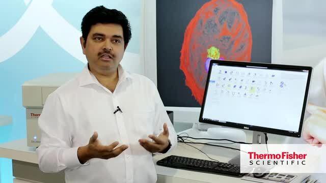
Thermo Fisher Cloud provides scientists with a secure place to store, share and analyze their data. Upload your files from anywhere you have access to the Internet by simply dragging them to the Cloud folder on your Windows™ desktop. Share the results with your colleagues to observe patterns, validate findings, and develop insights. And it’s not just for data files. Upload a presentation for an upcoming conference or a particularly useful how-to video.

Increase your screening throughput with CellInsight CX5 Platform
With the compact and powerful CellInsight CX5 High-Content Screening Platform you can read and analyze a 96-well plate in under four minutes. Designed for researchers looking to increase their screening throughput, the platform offers common excitation wavelengths for fluorescence and white light illumination for colorimetric samples. The system is ready for full automation with 96-, 384-, or 1,536-well plates, or you can analyze your tissue sections one slide at a time to work up your next assay.
CellInsight CX5 system highlights
| Illumination | 5-channel solid-state LED light engine |
| Camera | Photometrics High-Resolution Fluorescent Camera |
| Widefield | 5-channel fluorescent imaging |
| Brightfield | White light brightfield imaging |
| Objectives | Standard: 10x, 20x; Optional: 2x, 4x, 40x, 60x |
| Focus | Software autofocus |
| Automation | Configured for fully automated plate handling and scanning |
| Software | HCS Studio software for integrated data collection and analysis |

BPAE cells imaged using the CellInsight CX5 platform and stained with DAPI (blue), Alexa Fluor 568 phalloidin (red), and goat anti–mouse IgG (H+L) secondary antibody, Alexa Fluor 488 conjugate (green).

HeLa cells imaged using the CellInsight CX5 platform and stained with Hoechst 33342 (blue) and Alexa Fluor 680 phalloidin (yellow).

White light brightfield image of a fixed tissue slice stained with Hematoxylin and Eosin imaged with the CellInsight CX5 platform.
Reagents optimized for high-content screening
Drawing on decades of experience in fluorescence imaging, our HCS products are developed using Thermo Scientific High-Content Platforms with special considerations for the high-throughput workflow and automated imaging:
- Cell and nuclear masks for automated demarcation
- Robust functional probes for cell health interrogation
- Flexible assay workflow for automated processing
- Broad fluorophore choice for easy multiplexing
- Validated on multiple imaging platforms
Select reagents for use with the CellInsight CX5
Other cell structure and segmentation tools
Clear identification of cell and nuclear boundaries in automated imaging requires fiducial markers that will label cellular structures robustly and reproducibly. Cell masks are designed specifically for automated imaging. Molecular Probes cell structure labels are widely used in microscope imaging and HCS.
See other reagents for cell structure
Other cell health and cytotoxicity assays
We offer a diverse selection of imaging and microscopy assays for the analysis of cell viability, proliferation apoptosis, cell cycle and DNA repair, migration, adhesion and other cellular functions. Many of these assays have been validated specifically for HCS platforms.
Browse cell health and cytotoxicity assays by application:
Microplates and slides
Compatible with SBS standard microplates (6–1,536 well) and slides
Automation
Orbitor RS 2 Microplate Mover
Increase throughput, storage capacity and operational flexibility with the Thermo Scientific Orbitor RS 2 Microplate Mover—industrial-sized performance within a compact, benchtop mover.
The Orbitor RS 2 is a high-speed microplate mover offering proven, reliable performance with totally flexible plate handling. Extensive vertical reach allows multiple stacked or high density instruments to be loaded in a small footprint, and a bi-directional telescoping arm provides superior reach, improved user safety and unlimited base rotations within a 360° workspace.
- Learn more about the Orbitor RS 2 Microplate Mover
| Camera | Photometrics High-Resolution Fluorescent Camera:
|
| Data management | Compatible with Thermo Scientific Store Image and Database Management Software |
| Dimensions | Height: 37.2 cm (14.6 in) Width: 41.4 cm (16.3 in) Depth: 59.4 cm (23.4 in) |
| Image compatibility | JPG, BMP, GIF, PNG, TIF, C01, DIB |
| Light source | LED, solid-state five-color light engine used with provided filter sets offers the following excitation/emission capabilities (Ex/Em, in nm):
|
| Objectives | Standard (Olympus objectives):
|
| PC | Windows 10 Professional 64-Bit PC with 32GB (4x8GB) RAM, Intel™ Xeon™ Processor 3.7GHz Turbo, RAID I Controller with 1.8TB Hard Drive and 256 Boot Drive. Includes Dell 24" High Resolution Widescreen Monitor or equivalent, keyboard and mouse. |
| Software | Thermo Scientific HCS Studio 4.0 Cell Analysis Software |
| Wattage | 300 W |
| Weight | 45 kg (99.2 lb) |

Get started today with 10 GB of free storage.
Learn more
Access it now Use your existing web account username and password or register
Reagents optimized for high-content screening
Drawing on decades of experience in fluorescence imaging, our HCS products are developed using Thermo Scientific High-Content Platforms with special considerations for the high-throughput workflow and automated imaging:
- Cell and nuclear masks for automated demarcation
- Robust functional probes for cell health interrogation
- Flexible assay workflow for automated processing
- Broad fluorophore choice for easy multiplexing
- Validated on multiple imaging platforms
Select reagents for use with the CellInsight CX5
Other cell structure and segmentation tools
Clear identification of cell and nuclear boundaries in automated imaging requires fiducial markers that will label cellular structures robustly and reproducibly. Cell masks are designed specifically for automated imaging. Molecular Probes cell structure labels are widely used in microscope imaging and HCS.
See other reagents for cell structure
Other cell health and cytotoxicity assays
We offer a diverse selection of imaging and microscopy assays for the analysis of cell viability, proliferation apoptosis, cell cycle and DNA repair, migration, adhesion and other cellular functions. Many of these assays have been validated specifically for HCS platforms.
Browse cell health and cytotoxicity assays by application:
Microplates and slides
Compatible with SBS standard microplates (6–1,536 well) and slides
Automation
Orbitor RS 2 Microplate Mover
Increase throughput, storage capacity and operational flexibility with the Thermo Scientific Orbitor RS 2 Microplate Mover—industrial-sized performance within a compact, benchtop mover.
The Orbitor RS 2 is a high-speed microplate mover offering proven, reliable performance with totally flexible plate handling. Extensive vertical reach allows multiple stacked or high density instruments to be loaded in a small footprint, and a bi-directional telescoping arm provides superior reach, improved user safety and unlimited base rotations within a 360° workspace.
- Learn more about the Orbitor RS 2 Microplate Mover
| Camera | Photometrics High-Resolution Fluorescent Camera:
|
| Data management | Compatible with Thermo Scientific Store Image and Database Management Software |
| Dimensions | Height: 37.2 cm (14.6 in) Width: 41.4 cm (16.3 in) Depth: 59.4 cm (23.4 in) |
| Image compatibility | JPG, BMP, GIF, PNG, TIF, C01, DIB |
| Light source | LED, solid-state five-color light engine used with provided filter sets offers the following excitation/emission capabilities (Ex/Em, in nm):
|
| Objectives | Standard (Olympus objectives):
|
| PC | Windows 10 Professional 64-Bit PC with 32GB (4x8GB) RAM, Intel™ Xeon™ Processor 3.7GHz Turbo, RAID I Controller with 1.8TB Hard Drive and 256 Boot Drive. Includes Dell 24" High Resolution Widescreen Monitor or equivalent, keyboard and mouse. |
| Software | Thermo Scientific HCS Studio 4.0 Cell Analysis Software |
| Wattage | 300 W |
| Weight | 45 kg (99.2 lb) |

Thermo Fisher Cloud provides scientists with a secure place to store, share and analyze their data. Upload your files from anywhere you have access to the Internet by simply dragging them to the Cloud folder on your Windows™ desktop. Share the results with your colleagues to observe patterns, validate findings, and develop insights. And it’s not just for data files. Upload a presentation for an upcoming conference or a particularly useful how-to video.
Get started today with 10 GB of free storage.
Learn more
Access it now Use your existing web account username and password or register
Learn more about 2D/3D visualization and analysis (2D–5D)
With incredible speed and flexibility, Amira Software supports advanced 2D–5D bioimaging workflows in research areas ranging from structural and cellular biology to tissue imaging, neuroscience, preclinical imaging and bioengineering.
Software
Product brochures
Posters
Service
We provide a full range of service options for all EVOS cell imaging systems.
Application notes
Support
For Research Use Only. Not for use in diagnostic procedures.


