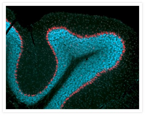
Neural somata in mouse cerebellum labeled with NeuroTrace® 530/615 red-fluorescent Nissl stain and DAPI.
Neural somata labeled with NeuroTrace® 530/615 red-fluorescent Nissl stain (Cat. no. N21482). Nuclei in this mouse cerebellum section were counterstained with DAPI (Cat. no. D1306, D3571, D21490). The image is a composite of two micrographs acquired using filters appropriate for tetramethylrhodamine and DAPI.

Related Products
Related Images
Developing Drosophila embryo Go ›

Cytoskeleton of a mixed population of granule neurons and glial cells Go ›

Cyanobacteria. BODIPY® FL, DAPI and CellTracker™ Green BODIPY®. Go ›

Immunofluorescent Staining using Mouse HuC/HuD Neuronal Protein Monoclonal Antibody, Biotin-XX Conjugate. Go ›

1% Agarose gel containing 16S and 23S ribosomal RNA (rRNA). SYBR® Green II RNA gel stain. Go ›

Bovine pulmonary artery endothelial cells (BPAEC). MitoTracker® Red CMXRos, SYTOX® Green nucleic acid stain, biotin-XX goat anti–mouse IgG antibody and Cascade Blue® NeutrAvidin biotin-binding protein. Go ›

CD335 (NKp46) Antibody (63335182) in RE Go ›

CD223 (LAG-3) Antibody (56223942) in TM Go ›
