Go further with your 3D life science data analysis

Thermo Scientific Amira Software is a powerful, multifaceted 3D platform for visualizing, manipulating, and understanding life science data from computed tomography, microscopy, MRI, and many other imaging modalities.
Amira Software is a configurable solution. Select the extensions that provide the best framework for your 3D data visualization and analysis needs.
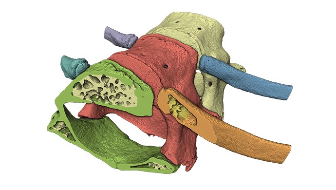
This extension provides advanced tools for image enhancement, simplified and automated segmentation, extensive measurement and quantification of objects, cells, and tissue features.

Courtesy of National Laboratory Animal Center - National Applied Research Laboratories, Taiwan
Conveniently load 3D or 4D+ data from over 150 file formats (powered by Bio-Formats, in cooperation with Glencoe Software).
This extension allows you to read data from the latest microscopes with the most current version of supported proprietary file formats.
Bioformats now integrates with XLVolume:
This extension allows detection and tracing of filaments or tube-like structures in very noisy images, enabling, for instance, analysis of actin fibers or microtubules in electron tomograms.

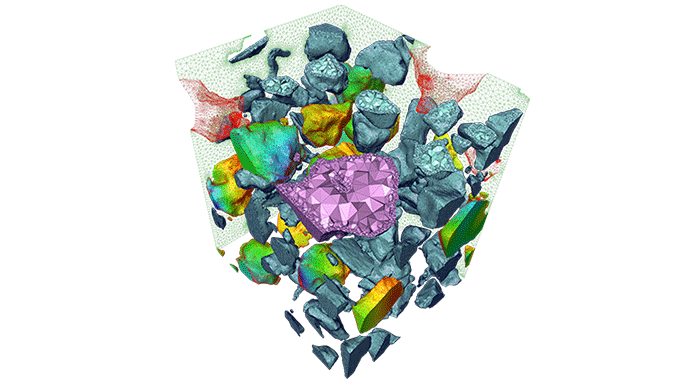
High-performance mesher allows for the generation of high-quality 3D tetrahedral mesh for FEA/CFD simulation.This extension is also dedicated to advanced post-processing of simulation data, ranging from flow to thermal, and stress data.

Manage and visualize very large amounts of volume data of up to several terabytes. Go far beyond the limit of the available system memory.
The multiresolution technique used in XLVolume extension enables interactive visualization and navigation through large data sets.
This extension combines Amira Software's strong capabilities for 3D data visualization, such as hardware-accelerated volume rendering, with specific tools for molecular visualization and analysis, such as molecular surfaces, sequence alignment, configuration density computation and molecule trajectory animation.

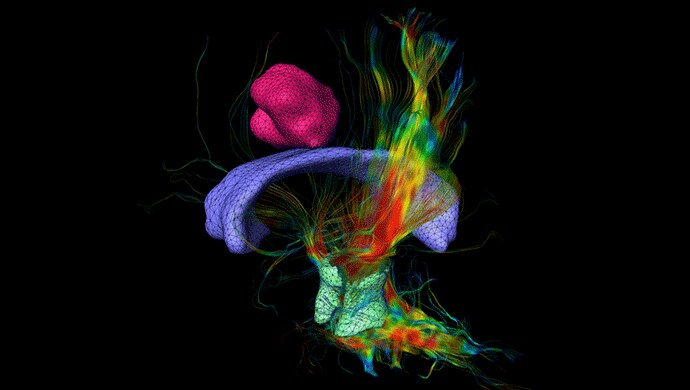
Amira Software solution to perform water diffusion analysis using diffusion weighted MRI. It enables the user to compute the apparent diffusion coefficient, fractional anisotropy, and directionally encoded color maps. Diffusion tensor imaging aims at localizing and visualizing fiber tracts. Full brain perfusion analysis can be performed on dynamic contrast enhanced MRI or CT studies.

This extension allows you to compute the precise internal displacement and strain maps from 3D images of objects acquired during a deformation process.

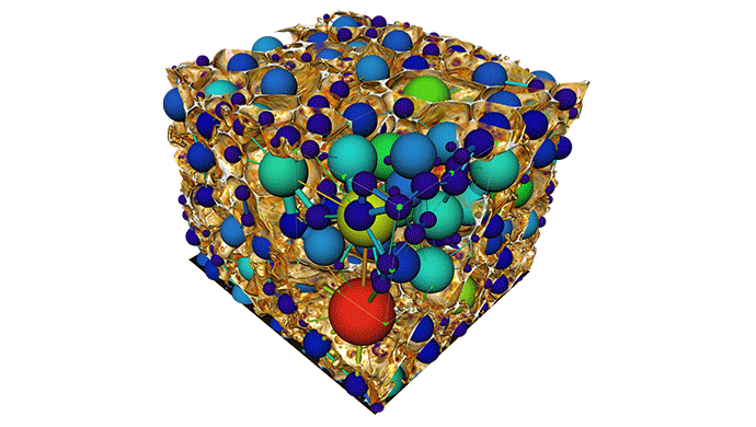
This extension allows for characterization and modeling of a set of connected areas, with spheres and lines representing those areas and their connections.

Create new custom components for Amira Software, such as file readers and writers, computation modules, and even new visualization modules, using the C++ programming language.