Studying protein function with cryo-EM
Cryo-EM is being applied to multiple scientific disciplines with great success, revealing molecular insights not achievable with any other technique. Highlighted below is just a fraction of the research being conducted with cryo-EM and where this technology can be applied with great advantages to synergize with other life science techniques.
Infectious disease research using cryo-electron microscopy
With an ever-increasing world population and global travel on the rise, infectious diseases cover more ground, spreading faster and more efficiently, leading to an increase in pandemic threats. Besides preventative (quarantine) measures, it is critical to combat diseases directly through the development of new antibiotics and/or efficient vaccines.
Particularly for viruses, determining the 3D outer shell structure can be vital, as that is where neutralizing antibodies interact with the pathogen. Cryo-EM techniques, such as single particle analysis, cryo-electron tomography (cryo-ET), and micro electron diffraction (MicroED), allow multiscale observation of these biological structures in their near-native states. With the full 3D structure, treatments can be motivated by observations on the molecular scale, greatly increasing specificity and overall success.
Our cryo-EM solutions are specially designed to deliver this vital information, leading to faster and more efficient development of therapeutics.
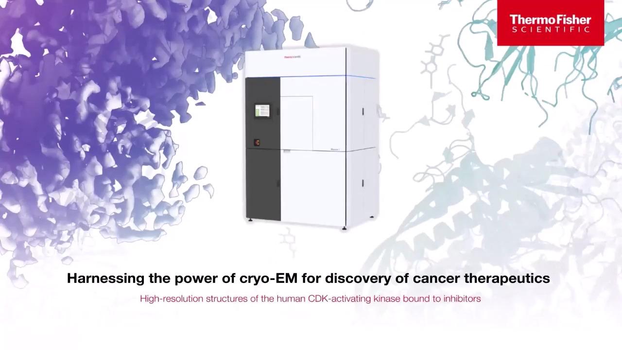
Breakthroughs in cancer research with cryo-EM
Genomic integrity and cell growth regulation lie at the heart of cancer. Proteins play a large role in mediating these mechanisms. Cryo-EM has become an invaluable tool in characterizing these protein structures and identifying how they behave during key cellular functions and mechanisms. Cryo-EM continues to revolutionize our understanding of cancer. Over the past several years, scientists have used cryo-EM to uncover new cancer pathways, enabling them to obtain an atomic-level description of key cancer protein targets and a framework for improving pharmaceutical interventions.
Neurodegenerative disease research using cryo-EM
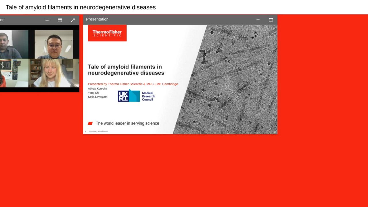
Neurodegenerative diseases encompass a broad range of neurological disorders characterized by the progressive dysfunction and degradation of neurons in the central nervous system. This includes well-known pathologies such as Alzheimer’s, Parkinson’s, and Huntington’s Disease as well as Amyotrophic Lateral Sclerosis (ALS). While the physical manifestation of these diseases has been extensively observed and characterized, the molecular basis is still an area of active investigation. By using structural biology, researchers have begun to unravel the molecular changes that lead to neurological disorder – these efforts, in turn, drive the development of therapeutics that can slow or stop the course of disease.
In recent years, cryo-EM technology has improved dramatically, enabling the characterization of large sample areas and macromolecular proteins. This is particularly important for neurodegenerative disease research, where disorders are often characterized by the agglomeration and precipitation of complex protein aggregates. Researchers have already utilized cryo-EM to uncover the atomic structures of numerous proteins associated with neurodegenerative diseases such as tau filaments, ɑ-synuclein fibrils, and amyloid ß aggregates, as well as small molecule drug candidates that bind to these structures.
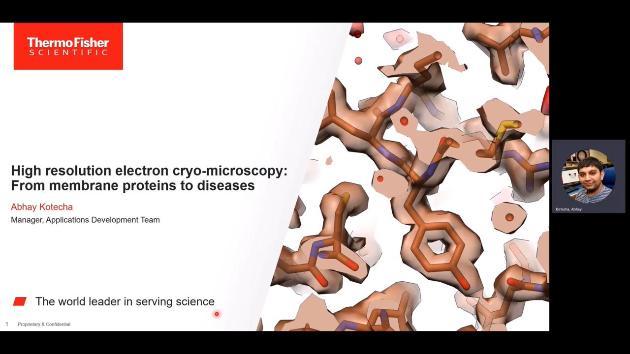
In general, protein structure and function correlate. By understanding the structural features of the protein aggregates implicated in neurodegenerative diseases, scientists can address how they form, interact with the cellular environment, and alter brain function. Membrane proteins and large macromolecular structures are challenging targets for X-ray crystallography and NMR spectroscopy (e.g., while membrane proteins account for over 60% of drug targets, they only make up ~2% of existing crystal structures). Cryo-EM techniques, meanwhile, do not require crystal growth, making them more flexible and capable of determining the structures of non-crystalline proteins. With cryo-EM, researchers can analyze the complex conformations, structures, and modified forms of proteins; multiple conformations can even be studied within a single sample.
Learn more about cryo-EM applications in neurodegenerative diseases
Biopharmaceutical research with electron microscopy
Cryo-EM techniques help examine a broad range of biological structures and provides the ability to visualize otherwise difficult or complicated biologics. For example, protein samples, which are frozen in vitreous (non-crystalline) ice, can be examined in a near-native, fully hydrated state, thereby enabling accelerated structural drug discovery. Near-atomic resolution cryo-electron microscopy can be employed throughout the drug discovery process, from target identification, through lead characterization, to epitope mapping.
Learn more on how cryo-EM is changing biopharmaceutical research
For Research Use Only. Not for use in diagnostic procedures.


