be51- Apps/Techniques/Products/Contact Us/Learning Center (LS)

Proteins serve as the primary molecular machinery of life, maintaining the functional order of our cells. As their structure informs their function, accurate determination of structural information is the basis of biological insights for any number of physiological, pharmaceutical, and biomaterial advances.

Cryo-electron microscopy (cryo-EM) is an established method for the determination of 3D structural information for a wide range of biomolecules, including proteins and protein complexes.
Until recently, proteins had to be crystallized to reconstruct and visualize them with X-ray crystallography. This information came with several stipulations; crystallization is time consuming and can typically only work on single purified protein (monomers or dimers); additionally, some proteins do not crystallize. Finally, the structure has to be determined outside of the cellular environment, removing vital contextual information.

Cryo-EM, which is comprised of several applications like single particle analysis (SPA), cryo-electron tomography (cryoET), and micro electron diffraction (MicroED), has revolutionized the study of proteins, their complexes, and their dynamics.

  • Single particle analysis (SPA)Images and analyzes small (<250kDa) and intermediate sized proteins (>250kDa) as well as large protein complexes with high throughput and high resolution.
  • Cryo tomography (cryoET)Ideal for the analysis of cellular components and fractions at intermediate resolutions. It provides cellular context to the macromolecular structures determined with single particle analysis.
  • Micro electron diffraction (MicroED)Suited for structural determination of small sub-750-nm protein crystals, a unique class of crystals that cannot be analyzed by traditional X-ray crystallography.
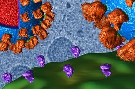
GABA receptor protein within a lipid nanodisk, generated with cryo electron microscopy.
3D reconstructions of the GABA receptor membrane protein in a nanodisk, bound to the drug Ro-15-4513. Displayed resolution is 2.75 Å. Data collected on a Krios Cryo-TEM with the Falcon 4 Detector. Images Courtesy of Simonas Masiulis, Radu Aricescu, MRC-LMB Cambridge and Evgenia Pechnikova, Abhay Kotecha, Thermo Fisher Scientific.
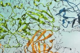
Granulin protomer structure generated by micro electron diffraction.
Granulin protomer (29 kDa) structure generated by cryo electron microscopy (MicroED) with atomic-resolution detail visible.
Electron Microscopy

Protein Electron Microscopy

Protein cryo-EM analysis for near-atomic structural insight.


Applications

Structural biology research

Structural Biology Research

Cryo-electron microscopy enables the structural analysis of challenging biological targets such as large complexes, flexible species and membrane protein.

Infectious disease research

Infectious Disease Research

Cryo-EM techniques enable multiscale observations of 3D biological structures in their near-native states, informing faster, more efficient development of therapeutics.

Drug discovery

Drug Discovery

Learn how to take advantage of rational drug design for many major drug target classes, leading to best-in-class drugs.


Techniques

Single Particle Analysis

Single particle analysis (SPA) is a cryo-electron microscopy technique that enables structural characterization at near-atomic resolutions, unraveling dynamic biological processes and the structure of biomolecular complexes/assemblies.

Learn more ›

Cryo-Tomography

Cryo-electron tomography (cryo-ET) delivers both structural information about individual proteins as well as their spatial arrangements within the cell. This makes it a truly unique technique and also explains why the method has such an enormous potential for cell biology. Cryo-ET can bridge the gap between light microscopy and near-atomic-resolution techniques like single-particle analysis.

Learn more ›

MicroED

MicroED is an exciting new technique with applications in the structural determination of small molecules and protein. With this method, atomic details can be extracted from individual nanocrystals (<200 nm in size), even in a heterogeneous mixture.

Learn more ›

Single Particle Analysis

Single particle analysis (SPA) is a cryo-electron microscopy technique that enables structural characterization at near-atomic resolutions, unraveling dynamic biological processes and the structure of biomolecular complexes/assemblies.

Learn more ›

Cryo-Tomography

Cryo-electron tomography (cryo-ET) delivers both structural information about individual proteins as well as their spatial arrangements within the cell. This makes it a truly unique technique and also explains why the method has such an enormous potential for cell biology. Cryo-ET can bridge the gap between light microscopy and near-atomic-resolution techniques like single-particle analysis.

Learn more ›

MicroED

MicroED is an exciting new technique with applications in the structural determination of small molecules and protein. With this method, atomic details can be extracted from individual nanocrystals (<200 nm in size), even in a heterogeneous mixture.

Learn more ›

Products

Style Sheet for Instrument Cards Original
Thermo Scientific Krios G4 Cryo transmission electron microscope (TEM)

Krios G4 Cryo-TEM

  • Improved ergonomics
  • Fits more easily into new and existing labs
  • Maximized productivity and automation
  • Best image quality for high-resolution 3D reconstruction
Thermo Scientific Glacios 2 Cryo-Transmission Electron Microscope

Glacios 2 Cryo-TEM

  • High-resolution structure determination tool (<3Å)
  • Unparalleled ease-of-use through AI-enhanced automation
  • Flexible modes of operation standard: SPA, tomography, MicroED
Thermo Scientific Tundra Cryo transmission electron microscope (TEM)

Tundra Cryo-TEM

  • Structural information at biologically relevant resolution
  • Space efficient and cost effective
  • Easy, iterative sample optimization
  • Unique AI algorithms for streamlined data collection
Thermo Scientific Talos F200C transmission electron microscope (TEM)

Talos F200C TEM

  • High-contrast and high-quality TEM and STEM imaging
  • 4k x 4k Ceta CMOS camera options for large FOV and high read-out speeds
  • Large pole piece gap and multiple in situ options
Thermo Scientific Aquilos 2 cryo focused ion beam (FIB)

Aquilos 2 Cryo-FIB

  • Automation enables production of multiple lamellas
  • Target and extract your structure of interest with lift-out nano-manipulator
  • 3D visualization for high-resolution tomography
Thermo Scientific Vitrobot vitrification sample system

Vitrobot System

  • Fully automated sample vitrification
  • Blotting device
  • Semi-automated grid transfer
  • High sample throughput
Thermo Scientific Selectris and Selectris X cryo electron microscopy imaging filters

Selectris and Selectris X Imaging Filters

  • Designed for high stability and atomic resolution imaging
  • Straightforward operation
  • Paired with the latest generation Thermo Scientific Falcon 4i Direct Electron Detector
Thermo Scientific Ceta-D camera for electron diffraction

Ceta-D Camera

  • Optimum performance at any high tension (20–300 kV)
  • Compatible with post-column filters and spectrometers
  • Movie acquisition for dynamic studies
Thermo Scientific Falcon 4i Electron Detector

Falcon 4i Detector

  • High throughput for more images per hour
  • Unsurpassed imaging quality with high DQE
  • Lossless data compression with EER
Thermo Scientific TEM Tomography 5 software

Tomography 5
Software

  • Automated multi-site batch tomography
  • Automatic cassette mapping
  • Dose symmetric tilt scheme for optimal electron dose
  • Fully integrated with Selectris Imaging Filters
Thermo Scientific Maps electron microscopy software

Maps Software

  • Acquire high-resolution images over large areas
  • Easily find regions of interest
  • Automate image acquisition process
  • Correlate data from different sources
Thermo Scientific Auto Slice and View 4.0 serial section electron microscopy software

Auto Slice and View 4.0 Software

  • Automated serial sectioning for DualBeam
  • Multi-modal data acquisition (SEM, EDS, EBSD)
  • On-the-fly editing capabilities
  • Edge based cut placement
Thermo Scientific Inspect 3D tomography software

Inspect 3D Software

  • Image processing tools and filters for cross-correlation
  • Feature tracking for image alignment
  • Algebraic reconstruction technique for iterative projection comparison
Thermo Scientific Amira 2D-5D visualization Software

Amira Software
Life Sciences

  • Explore 2D-5D bioimaging data
  • Identify and understand structures
  • Obtain statistical information
  • Share reports and stunning animations
Thermo Scientific EPU Software for single particle analysis

Smart EPU Software

  • Microscope-embedded solution for single particle acquisition
  • Optimized for high-throughput particle collection, providing real-time image analysis
  • Includes components for scheduling, data acquisition, and decision algorithms

Contact us

Style Sheet to change H2 style to p with em-h2-header class
Style Sheet to change H3 to p with em-h3-header class
Style Sheet to change Applications H3 to p with em-h3-header class
Style Sheet for Komodo Tabs

Electron microscopy services for
the life sciences

To ensure optimal system performance, we provide you access to a world-class network of field service experts, technical support, and certified spare parts.

Style Sheet for Support and Service footer
Style Sheet for Fonts
Style Sheet for Cards