Invitrogen
Phospho-MCL-1 (Ser159) Monoclonal Antibody (RBCERNR), PE, eBioscience™
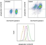
FIGURE: 1 / 1
Phospho-MCL-1 (Ser159) Antibody (12-9038-42) in Flow

Product Details
12-9038-42
Species Reactivity
Host/Isotype
Recommended Isotype Control
Class
Type
Clone
Conjugate
Excitation/Emission Max
Form
Concentration
Purification
Storage buffer
Contains
Storage conditions
Shipping conditions
RRID
Product Specific Information
Description: This RBCERNR monoclonal antibody recognizes human and mouse myeloid cell leukemia sequence 1 (Mcl-1) when phosphorylated on serine 159 (S159). Mcl-1 is an anti-apoptotic protein that is a member of the Bcl-2 family of proteins important for regulation of cell survival/apoptosis. Mcl-1 is primarily localized to the outer membrane of mitochondria where it prevents cytochrome c release via dimerization with other Bcl-2 family members such as Bim. PI3K activation of AKT results in the phosphorylation of GSK3 beta at serine 9 (S9) resulting in destabilization and degradation of GSK3 beta. Loss of GSK3 beta prevents phosphorylation of Mcl-1 on S159 and its subsequent ubiquitnation and degradation. Mice conditionally lacking Mcl-1 in lymphocytes showed that Mcl-1 is essential during early lymphoid development and for the maintenance of mature lymphocytes.
Applications Reported: This RBCERNR antibody has been reported for use in intracellular staining followed by flow cytometric analysis.
Applications Tested: This RBCERNR antibody has been pre-titrated and tested by intracellular staining followed by flow cytometric analysis of normal human peripheral blood cells. This can be used at 5 µL (0.125 µg) per test. A test is defined as the amount (µg) of antibody that will stain a cell sample in a final volume of 100 µL. Cell number should be determined empirically but can range from 10^5 to 10^8 cells/test.
Staining Protocol: Protocol A and Protocol C are recommended for this monoclonal antibody. Use of Protocol A: Two-step protocol: intracellular (cytoplasmic) proteins allows for the greatest flexibility for detection of surface and intracellular (cytoplasmic) proteins. Protocol C: Two-step protocol: Fixation/Methanol allows for the greatest discrimination of phospho-specific signaling between unstimulated and stimulated samples, but with limitations on the ability to stain specific surface proteins (refer to "Clone Performance Following Fixation/Permeabilization" located in the Best Protocols Section under the Resources tab online). All Protocols can be found in the Flow Cytometry Protocols: "Staining Intracellular Antigens for Flow Cytometry Protocol" located in the Best Protocols Section under the Resources tab online.
Excitation: 488-561 nm; Emission: 578 nm; Laser: Blue Laser, Green Laser, Yellow-Green Laser.
Filtration: 0.2 µm post-manufacturing filtered.
Target Information
MCL1 (Myeloid cell leukemia-1) belongs to the Bcl-2 family and is involved in programing, differentiation and concomitant maintenance of cell viability, but not of proliferation. Isoform 1 of MCL1 inhibits apoptosis while isoform 2 promotes it. The carboxy terminal of MCL1 and bcl-2 share significant sequence homology. Expression of MCL1 is increased upon exposure of ML-1 cells to various types of DNA damaging agents (e.g. ionizing radiation, ultraviolet radiation, and alkylating drugs) along with increases in GADD45 and Bax and a decrease in bcl-2. Enhanced expression of MCL1, prominently associated with mitochondria, complements the continued expression of bcl-2 in ML-1 cells undergoing differentiation. Like bcl-2, MCL1 has the capacity to promote cell viability under conditions that otherwise cause apoptosis. While the mechanism by which MCL1 inhibits apoptosis is not known, it is thought that it may heterodimerize and neutralize pro-apoptotic members of the Bcl-2 family such as Bim or Bak. MCL1 was originally identified in differentiating myeloid cells, but has since been shown to be expressed in multiple cell types. MCL1 is essential for embryogenesis and for the development and maintenance of B and T lymphocytes in animals. MCL1 exists as at least two distinct isoforms designated MCL1L and MCL1S. In marked contrast to the larger isoform of MCL1, overexpression of MCL1S promotes cell death.
For Research Use Only. Not for use in diagnostic procedures. Not for resale without express authorization.
How to use the Panel Builder
Watch the video to learn how to use the Invitrogen Flow Cytometry Panel Builder to build your next flow cytometry panel in 5 easy steps.
References (0)
Bioinformatics
Protein Aliases: Bcl-2-like protein 3; Bcl-2-related protein EAT/mcl1; Bcl2-L-3; Induced myeloid leukemia cell differentiation protein Mcl-1; Induced myeloid leukemia cell differentiation protein Mcl-1 homolog; MCL-1S; mcl1/EAT; MGC104264; MGC1839; myeloid cell leukemia 1; myeloid cell leukemia ES; myeloid cell leukemia sequence 1 (BCL2-related)
Gene Aliases: AW556805; bcl2-L-3; BCL2L3; EAT; Mcl-1; MCL1; MCL1-ES; mcl1/EAT; MCL1L; MCL1S; TM
UniProt ID: (Human) D3DV04, (Mouse) P97287
Entrez Gene ID: (Human) 4170, (Mouse) 17210

Performance Guarantee
If an Invitrogen™ antibody doesn't perform as described on our website or datasheet,we'll replace the product at no cost to you, or provide you with a credit for a future purchase.*
Learn more
We're here to help
Get expert recommendations for common problems or connect directly with an on staff expert for technical assistance related to applications, equipment and general product use.
Contact tech support

