As materials continue to advance, it is becoming increasingly important to not only examine them at ever-higher resolutions but to obtain these observations within the relevant macroscopic context. This necessitates correlating different imaging modes to the same coordinates for truly contextual insight. Measurements must also be obtained quickly enough for practical application in real-world process control and failure analysis environments. Thermo Fisher Scientific offers a complete workflow for the observation of materials, combining correlated imaging at various scales with additional information such as chemical composition.
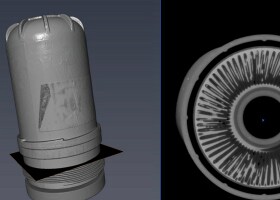
Multi-scale analysis begins with micro-scale observation with non-destructive spectroscopic techniques. X-ray microtomography (microCT) produces a complete, 3D rendering of the sample through serial X-ray scans. These scans, or 2D tomograms, are digitally combined to form the 3D structure. With Thermo Scientific Heliscan MicroCT, the series of circular scans is replaced by a single, continuous helical scan. This allows for faster scanning at a lower dose, increasing the accuracy and amount of information obtained. MicroCT observations can provide resolution as low as 400 nm, making it an ideal tool for non-destructive surveying of the sample prior to higher resolution characterization.
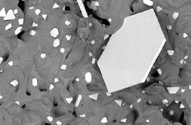
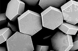
Once a region of interest is identified, DualBeam (focused ion beam and scanning electron microscopy, FIB-SEM) instrumentation is used for closer surface analysis and sample extraction. (Note that the focused ion beam can consist of a liquid metal ion source (gallium) or a plasma FIB.) The SEM enables nanoscale surface analysis while the FIB/PFIB is used for serial sectioning, or to extract a thin sample lamella for further observation with transmission electron microscopy (TEM). The addition of a femtosecond laser to the PFIB-SEM allows for even more rapid sample preparation, cross-sectioning or serial sectioning. Subsequent TEM analysis provides atomic-scale materials characterization for complete insight into a sample’s elemental and structural composition.
True multi-scale microscopy generates high quality and reliable imaging across all instruments while also accurately aligning them into a complete representation of the sample. With Thermo Scientific automation and data analysis software, the entire multi-scale workflow becomes a guided and routine procedure that can be readily integrated into your process or quality control environment.


