Invitrogen
Phospho-Histone H2A.X (Ser139) Recombinant Mouse Monoclonal Antibody (CR55T33), Alexa Fluor™ Plus 555
This Antibody was verified by Cell treatment to ensure that the antibody binds to the antigen stated.
FIGURE: 1 / 3
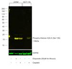
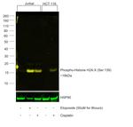
Phospho-Histone H2A.X (Ser139) Antibody (740078MP555) in WB


Product Details
740078MP555
Species Reactivity
Host/Isotype
Expression System
Class
Type
Clone
Conjugate
Excitation/Emission Max
Form
Concentration
Purification
Storage buffer
Contains
Storage conditions
Shipping conditions
Product Specific Information
Alexa Fluor™ Plus recombinant antibodies are conjugated using new, proprietary dye chemistry so you can generate stunning data. Alexa Fluor™ Plus antibodies represent an advancement in fluorescent conjugate technology. Alexa Fluor™ Plus antibodies provide brighter signal compared to leading Alexa Fluor™ antibodies, providing you with better signal-to-noise for your critical experiments. These antibodies show better specificity and lot-to-lot consistency as these are recombinant antibodies, generated by cloning specific genes for the desired antibodies into an expression vector and expressed in vitro.
Using conjugate solutions: Centrifuge the protein conjugate solution briefly in a microcentrifuge before use; add only the supernatant to the experiment. This step will help eliminate any protein aggregates that may have formed during storage, thereby reducing nonspecific background staining.
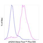
Applications Tested: This 740078MP555 antibody (CR55T33) has been tested by western blot and Flowcytometry analysis of Jurkat cells treated with 50µM etiposide for 8 hrs. This may be used for western blotting at 2 µg/ml and for western blotting. It is recommended that the antibody be carefully titrated for optimal performance in the assay of interest.
Excitation: 553 nm; Emission: 568 nm; Laser: Yellow Laser
Filtration: 0.2 µm post-manufacturing filtered.
Target Information
Histone H2A.X (H2AX) is a member of the histone H2A family which is one of the four core histones making up the nucleosome core particle. In eukaryotes, DNA double strand breaks (DSBs) have been shown to trigger the phosphorylation of serine 139 at the carboxy terminus of histone H2AX resulting in gamma-H2AX. The phosphorylation of H2AX can be detected by Western blotting or immunofluorescence, revealing the frequency of DSBs. The phosphatidylinositol 3-kinases have been implicated in H2AX phosphorylation, but it is unclear if ATM is the primary H2AX kinase or if other members of the family such as DNA-PK and ATR contribute in a similar manner. Structurally, H2A.x contains 143 amino acid residues. Histone H2A.X is considered a basal histone, being synthesized in G1 as well as in S-phase, and its mRNA contains polyA addition motifs and a polyA tail along with the conserved stem-loop and U7 binding sequences involved in the processing and stability of replication type histone mRNAs. There are two forms of Histone H2A.X mRNA, one about 1600 bases long and contains polyA; the other about 575 bases long, lacking polyA. The short form behaves as a replication type histone mRNA, while the longer behaves as a basal type histone mRNA. Histone H2A.X maps to the 11q23.2-q23.3 region of the human chromosome. Histone H2A.x contributes to histone-formation and therefore the structure of DNA. Histone H2A variant H2A.x specifically regulates the interaction of MDC1 (mediator of DNA damage checkpoint protein 1), a DNA repair protein to the sites of DNA damage.
For Research Use Only. Not for use in diagnostic procedures. Not for resale without express authorization.
References (0)
Bioinformatics
Protein Aliases: gamma H2AX; H2A histone family, member X; H2a/x; H2AX histone; Histone H2A.X; Histone H2a/x; Histone H2AX
Gene Aliases: H2A.X; H2A/X; H2AFX; H2AX
UniProt ID: (Human) P16104
Entrez Gene ID: (Human) 3014

Performance Guarantee
If an Invitrogen™ antibody doesn't perform as described on our website or datasheet,we'll replace the product at no cost to you, or provide you with a credit for a future purchase.*
Learn more
We're here to help
Get expert recommendations for common problems or connect directly with an on staff expert for technical assistance related to applications, equipment and general product use.
Contact tech support

