Invitrogen
MERTK Monoclonal Antibody (DS5MMER), Alexa Fluor™ 488, eBioscience™
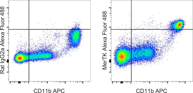
FIGURE: 1 / 7
MERTK Antibody (53-5751-82) in Flow


Product Details
53-5751-82
Species Reactivity
Published species
Host/Isotype
Recommended Isotype Control
Class
Type
Clone
Immunogen
Conjugate
Excitation/Emission Max
Form
Concentration
Purification
Storage buffer
Contains
Storage conditions
Shipping conditions
RRID
Product Specific Information
Description: This DS5MMER monoclonal antibody recognizes mouse MerTK, a 170-210 kDa member of the TAM family of tyrosine kinase receptors that also includes Axl and Tyro3. MerTK is expressed on tissue macrophages and is involved in the removal of apoptotic cells. This process relies on two soluble ligands of MerTK, Protein S and Gas6 that bind to phosphatidylserine found on the outer leaflet of the plasma membrane of cells undergoing apoptosis. Upon binding these ligands, MerTK undergoes autophosphorylation at multiple tyrosine residues that activate the PI3K and Akt pathways. This results in the phagocytosis of apoptotic cells and also results in the direct inhibition of TLR-induced production of pro-inflammatory cytokines. In addition, MerTK may function as a putative entry receptor for filoviruses. Deficiency of MerTK causes general autoimmunity, inflammation and accumulation of apoptotic bodies. MerTK is constitutively released from the cell surface by metalloproteinases and thus is present in the serum and culture medium. This process can be enhanced by stimulation with LPS. MerTK can be used to help discriminate macrophages from dendritic cells. MerTK is often expressed on malignant cells and may be implicated in immune evasion.
The DS5MMER antibody will work on paraformaldehyde-fixed cells.
Applications Reported: This DS5MMER antibody has been reported for use in flow cytometric analysis.
Applications Tested: This DS5MMER antibody has been tested by flow cytometric analysis of mouse resident peritoneal cells. This may be used at less than or equal to 1.0 µg per test. A test is defined as the amount (µg) of antibody that will stain a cell sample in a final volume of 100 µL. Cell number should be determined empirically but can range from 10^5 to 10^8 cells/test. It is recommended that the antibody be carefully titrated for optimal performance in the assay of interest.
Excitation: 488 nm; Emission: 519 nm; Laser: Blue Laser
Target Information
MERTK (cMER) is a tyrosine kinase proto-oncogene and is involved in the retinal pigment epithelium (RPE) phagocytosis pathway, which is implicated in human retinal disease. Regulates many physiological processes including cell survival, migration, differentiation, and phagocytosis of apoptotic cells (efferocytosis). Ligand binding at the cell surface induces autophosphorylation of MERTK on its intracellular domain that provides docking sites for downstream signaling molecules. Following activation by ligand, interacts with GRB2 or PLCG2 and induces phosphorylation of MAPK1, MAPK2, FAK/PTK2 or RAC1. MERTK signaling plays a role in various processes such as macrophage clearance of apoptotic cells, platelet aggregation, cytoskeleton reorganization and engulfment. Functions in the retinal pigment epithelium (RPE) as a regulator of rod outer segments fragments phagocytosis. Plays also an important role in inhibition of Toll-like receptors (TLRs)-mediated innate immune response by activating STAT1, which selectively induces production of suppressors of cytokine signaling SOCS1 and SOCS3.
For Research Use Only. Not for use in diagnostic procedures. Not for resale without express authorization.
How to use the Panel Builder
Watch the video to learn how to use the Invitrogen Flow Cytometry Panel Builder to build your next flow cytometry panel in 5 easy steps.
Bioinformatics
Protein Aliases: MGC133349; Proto-oncogene c-Mer; proto-oncogene tyrosine-protein kinase Mer; Receptor tyrosine kinase MerTK; sMER; sMERTK; soluble MER; soluble MERTK; Tyro 12; Tyrosine-protein kinase Mer
Gene Aliases: Eyk; Mer; Mertk; nmf12; Nyk
UniProt ID: (Mouse) Q60805
Entrez Gene ID: (Mouse) 17289

Performance Guarantee
If an Invitrogen™ antibody doesn't perform as described on our website or datasheet,we'll replace the product at no cost to you, or provide you with a credit for a future purchase.*
Learn more
We're here to help
Get expert recommendations for common problems or connect directly with an on staff expert for technical assistance related to applications, equipment and general product use.
Contact tech support

