Invitrogen
F4/80 Monoclonal Antibody (BM8), Brilliant Violet™ 650, eBioscience™
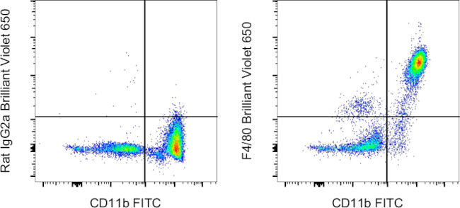
FIGURE: 1 / 1
F4/80 Antibody (416-4801-82) in Flow

Product Details
416-4801-82
Species Reactivity
Host/Isotype
Recommended Isotype Control
Class
Type
Clone
Conjugate
Excitation/Emission Max
Form
Concentration
Purification
Storage buffer
Contains
Storage conditions
Shipping conditions
RRID
Product Specific Information
Description: The BM8 monoclonal antibody reacts with mouse F4/80 antigen, an approximately 160 kDa surface receptor. It belongs to the EGF-TM7 family of proteins. As such it contains seven EGF-like domains on its extracellular N-terminus, seven transmembrane spanning sequences, and an intracellular C-terminal domain showing homology to other TM7 superfamily members. The F4/80 antigen is expressed by a majority of mature macrophages and is one of the best markers for this population of cells. However, other cell types, such as peritoneal eosinophils, Langerhans cells, and some other dendritic cell subtypes, have been reported to express this antigen as well. Expression of F4/80 commences during early myeloid development in vivo and can be upregulated on BM cells stimulated in vitro with M-CSF. Some populations of macrophages, especially in the lymphoid microenvironment, may be devoid of F4/80.
Applications Reported: This BM8 antibody has been reported for use in flow cytometric analysis.
Applications Tested: This BM8 antibody has been tested by flow cytometric analysis of mouse resident peritoneal macrophages. This may be used at less than or equal to 1.0 µg per test. A test is defined as the amount (µg) of antibody that will stain a cell sample in a final volume of 100 µL. Cell number should be determined empirically but can range from 10^5 to 10^8 cells/test. It is recommended that the antibody be carefully titrated for optimal performance in the assay of interest.
Brilliant Violet™ 650 (BV650) is a tandem dye that emits at 649 nm and is intended for use on cytometers equipped with a violet (405 nm) laser. Please make sure that your instrument is capable of detecting this fluorochrome.
When using two or more Super Bright, Brilliant Violet™, Brilliant Ultra Violet™, or other polymer dye-conjugated antibodies in a staining panel, it is recommended to use Super Bright Complete Staining Buffer (Product # SB-4401-42) or Brilliant Stain Buffer™ (Product # 00-4409-75) to minimize any non-specific polymer interactions. Please refer to the datasheet for Super Bright Staining Buffer or Brilliant Stain Buffer for more information.
Light sensitivity: This tandem dye is sensitive to photo-induced oxidation. Please protect this vial and stained samples from light.
Fixation: Samples can be stored in IC Fixation Buffer (Product # 00-8222-49) (100 µL of cell sample + 100 µL of IC Fixation Buffer) or 1-step Fix/Lyse Solution (Product # 00-5333-54) for up to 3 days in the dark at 4°C with minimal impact on brightness and FRET efficiency/compensation. Some generalizations regarding fluorophore performance after fixation can be made, but clone-specific performance should be determined empirically.
Our internal testing suggests that Brilliant Violet™ 650 (BV650) is not compatible with methanol-based fixation.
Excitation: 407 nm; Emission: 649 nm; Laser: Violet Laser.
BRILLIANT VIOLET™ is a trademark or registered trademark of Becton, Dickinson and Company or its affiliates, and is used under license. Powered by Sirigen™.
Target Information
F4/80 antigen is a 160 kDa cell surface glycoprotein that is a member of the EGF TM7 family of proteins and shares 68% overall amino acid identity with human EGF module containing mucin like hormone receptor 1 (EMR1). Expression of F4/80 is heterogeneous and is reported to vary during macrophage maturation and activation. The F4/80 antigen is expressed on a wide range of mature tissue macrophages including Kupffer cells, Langerhans, microglia, macrophages located in the gut lamina propria, peritoneal cavity, lung, thymus, bone marrow stroma and macrophages in the red pulp of the spleen. F4/80 expression has also been reported on a subpopulation of dendritic cells but is absent from macrophages located in T cell areas of the spleen and lymph node. The ligands and biological functions of the F4/80 antigen have not yet been determined but recent studies suggest a role for F4/80 in the generation of efferent CD8+ve regulatory T cells.
For Research Use Only. Not for use in diagnostic procedures. Not for resale without express authorization.
How to use the Panel Builder
Watch the video to learn how to use the Invitrogen Flow Cytometry Panel Builder to build your next flow cytometry panel in 5 easy steps.
References (0)
Bioinformatics
Protein Aliases: Adhesion G protein-coupled receptor E1; Cell surface glycoprotein F4/80; EGF-like module containing, mucin-like, hormone receptor-like sequence 1; EGF-like module receptor 1; EGF-like module-containing mucin-like hormone receptor-like 1; EMR1 hormone receptor; lymphocyte antigen 71
Gene Aliases: Adgre1; DD7A5-7; EGF-TM7; Emr1; F4/80; Gpf480; Ly71; TM7LN3
UniProt ID: (Mouse) Q61549
Entrez Gene ID: (Mouse) 13733

Performance Guarantee
If an Invitrogen™ antibody doesn't perform as described on our website or datasheet,we'll replace the product at no cost to you, or provide you with a credit for a future purchase.*
Learn more
We're here to help
Get expert recommendations for common problems or connect directly with an on staff expert for technical assistance related to applications, equipment and general product use.
Contact tech support

